
Breathing Series, Part 2 of 4
Introduction
The previous part took up how breathing is carried out within the body, in its two forms — unconscious and conscious (see “Exhaling Takes No Effort — Conscious and Unconscious Breath“). It closed by noting that the muscles involved in inhalation are Tonic muscle, energy-efficient within the body and involved in maintaining posture, while the muscles involved in exhalation — the exhalation of forced breathing — fall under Phasic muscle.

This part addresses the diaphragm, one of the two principal muscles involved in breathing.
The Diaphragm and Breathing
The diaphragm sits on the boundary between the thoracic cavity and the abdominal cavity. Because it breathes by moving bone, it attaches to bone. Anatomy defines the pulling side — the part that does not move and serves as the fulcrum — as the origin, and the side that is pulled — the part that moves and serves as the point of action — as the attachment. Seen this way, the origins of the diaphragm lie at the xiphoid process, the ribs, and the lumbar vertebrae; the attachment lies at the central tendon.
When the signal for inhalation arrives from the respiratory center and the diaphragm at the lower ribs contracts, the central tendon is drawn downward. Pressure within the thoracic cavity falls, and air enters the lungs. As the central tendon descends, the abdominal cavity is pressed downward.
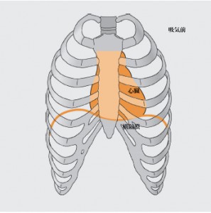

Within the abdominal cavity there are two sacs. The first is the peritoneal cavity, covered by the translucent peritoneum, which houses organs such as the liver and stomach. The second — called the subperitoneal cavity, meaning below the peritoneal cavity — contains the pelvic organs near the pelvic floor: the rectum, the uterus, the prostate, the bladder, and others.
Pressure from the thoracic cavity has a favorable effect on the former, while the latter requires protection from that same pressure. Ordinarily, when pressure from the diaphragm reaches the abdominal cavity, the pelvis tilts forward. The peritoneal cavity is pushed forward, and as a result the subperitoneal cavity and the pelvic organs descend, escaping the pressure from the diaphragm.

As an aside, the contraction of the diaphragm moves the viscera within the abdominal cavity. The liver and stomach are said to descend toward the navel, and the kidneys to move downward.
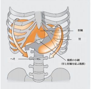

Thoracic Breathing and Abdominal Breathing
Because the organs move in this way, they come under the influence of the autonomic nerve fibers that supply them. Consider the relation among thoracic breathing, abdominal breathing, and the autonomic nervous system. Thoracic breathing works mainly through the spreading of the ribs, which widens the rib cage; abdominal breathing works by using the diaphragm while keeping the rib cage as still as possible. Thoracic breathing activates the sympathetic nervous system, abdominal breathing the parasympathetic — and out of that relation comes the attention abdominal breathing receives. In practice, breathing never occurs through one alone; the two work together.
One thing learned in the anatomy of Rolfing Phase I is that although a muscle has an origin and an attachment, either can become the origin of movement — in English, the Fixed Point. Personal habit and the relation to the environment (is this a safe environment, or one calling for vigilance?) alter posture. As posture alters the Fixed Point of a muscle, the manner of breathing changes with it.
Consider this from the environment in which the diaphragm sits. The diaphragm lies on the boundary between the abdominal and thoracic cavities, but seen from the body as a whole, it is understood to connect, through connective tissue, with the skull, the spine, and the heart.

In more detail: through connective tissue, the diaphragm forms a network with the heart and the spine — the portion connecting toward the spine is called the crura. And the heart connects to the clavicles and the skull by way of the anterior mediastinal ligament, the mediastinum being the middle of the thoracic cavity.

From this it follows that when the head tilts forward at a computer or a smartphone (pulled by the anterior mediastinal ligament), or when the curvature of the lumbar and thoracic spine changes (pulled by the crura), the movement of the central tendon narrows, and breathing is affected. Breathing, in turn, affects the placement of the organs.
On posture and breathing, the next part will take up the scalenes and the balance of extensors and flexors.
References
- Aline Newton; Breathing in the gravity field, Fall 1997, Rolf Lines
- Peter Schwind; Fascial and Membrane Technique, 2006
Note, August 2026
This piece was also written in February 2015, during the Phase III training. The terminology and internal references were put in order, and the whole was re-edited as a four-part series.
Breathing Series (Four Parts)
- Part 1: Exhaling Takes No Effort — Conscious and Unconscious Breath
- Part 2: The Diaphragm Reaches the Skull — Breath and the Placement of the Organs (this article)
- Part 3: When Posture Collapses, Breathing Changes — Extensors, Flexors, and the Scalenes
- Part 4: The Inhaling Body and the Exhaling Body — Breath Seen Through Gravity
This series was written during basic training. The same subject is taken up again eight years later, from the side of the research.
Breathing, Revisited (Four Parts)
- Part 1: A Body That Breathes Too Much — When Carbon Dioxide Runs Short
- Part 2: The Diaphragm Reaches the Heart — Breath, Heart Rate, and Two Nuclei in the Brainstem
- Part 3: The Jaw Is Made by Breathing — Mouth Breathing and the Teeth
- Part 4: There Are Sensors on the Inside — The Vagus Nerve and Interoception