
Breathing, Revisited — Part 2 of 4
This is the second of four pieces in the series Breathing, Revisited. The series returns to breathing from the side of the research, eight years into offering Rolfing sessions. It stands in a different place from the four-part breathing series written back in 2015, during basic training. This second piece follows the diaphragm along its connections to the heart and to the brain.
Breathing and the diaphragm
When I begin a first session, I ask:
“When you breathe, which is harder — breathing in, or breathing out?”
And I direct attention to the shoulders, the front and back of the chest, the sides of the ribcage.
The question is also asking something else: how much of the breathing musculature is in use?
In breathing, depth comes from the movement of the diaphragm and of the intercostals, the muscles between the ribs. Other muscles are involved, but I will keep to these two here. The first Rolfing session is built so that the movement of the diaphragm, above all, can be felt.
Bringing order to the diaphragm makes breathing easier to feel — and there is more to it than that.
- The diaphragm can influence heart rate.
- The diaphragm moves on a signal from the brain, and that signal can itself be influenced.
- A breathing practice — box breathing — can substantially improve the signal from the brain to the diaphragm.
What follows sets out what improved diaphragmatic movement brings.
Attending to the in-breath and the out-breath — awareness of breath can change heart rate
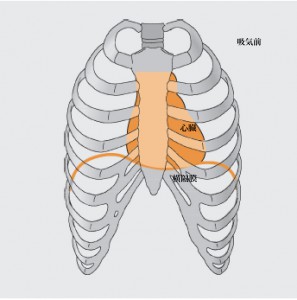
The diaphragm is a dome-shaped muscle beneath the ribs, dividing the trunk into the thorax, which holds the lungs and the heart, and the abdomen, which holds the intestines, the liver, and the other organs. Breathe in, and air enters the lungs; that pressure lowers the diaphragm. Breathe out, and air leaves the lungs; the diaphragm rises.

Breathing exerts considerable influence on the mind.
To concentrate, lengthen the in-breath; to relax, lengthen the out-breath.
The reason:
Breathing in raises heart rate, as the sympathetic branch of the autonomic nervous system becomes active. Breathing out lowers it, as the parasympathetic branch does.
In this way the movement of breath — the movement of the diaphragm — bears strongly on heart rate, and so on the mind.
The principle runs as follows.
Breathe in, and the diaphragm descends. The diaphragm is joined to the heart through fascia, so when the diaphragm moves, the heart moves with it. As the diaphragm descends, the heart is drawn downward and opens slightly. Blood is flowing within the heart, and being drawn on in this way slows that flow. From the sinoatrial node, which serves as the heart’s pacemaker, word goes to the brain:
“The flow of blood has slowed.”
On that information, the brain replies:
“Speed the flow of blood.”
That is, it signals the heart to raise the rate, and the rate rises. Breathing in raises heart rate.
Breathe out, and the diaphragm rises; the heart narrows slightly.
“The flow of blood has quickened.”
Word goes to the brain, and on that information the brain replies:
“Slow the flow of blood.”
It signals the heart to lower the rate, and the rate falls. Breathing out lowers heart rate.
Because we are breathing, the heart rate necessarily varies. The pulse slows and quickens. One measure of this variation is HRV — heart rate variability. HRV offers a reading of the state of the autonomic nervous system, and it has drawn attention in the world of athletics.
High HRV corresponds to parasympathetic predominance, low HRV to sympathetic predominance; that is the simplest way to hold it. Apps for phones are available, if you are curious.
The diaphragm moves on a signal from the brain — which nervous system is at work?
The muscles we can move at will — voluntary muscles — are skeletal, and they move on signals from the brain. The organs, the heart among them, are smooth muscle, governed by the autonomic nervous system and not movable at will: involuntary muscle. The diaphragm, for its part, moves on signals from the brain.
In breathing, the part of the brain involved is the brainstem.
Two sites there are known to take part in breathing.
- The pre-Bötzinger complex (preBötC), in the reticular formation of the ventrolateral medulla, around the back of the head near the neck
- The parafacial nucleus (PFN), in the retrotrapezoid nucleus of the ventrolateral medulla
The first was identified by Jack Feldman and colleagues at the University of California, Los Angeles, in 1991. It was named for the Bötzinger complex lying just beside it — and that name itself comes from a Bötzinger wine served at the dinner of a conference held in Germany in 1980.
The second was described in neonatal rat preparations as neurons below the facial motor nucleus showing biphasic pre-inspiratory discharge. That report dates from 2003, though the cell group itself goes back to work published in 1987.
The preBötC governs the rhythm of breathing — in, out, in — and works while we are asleep, or whenever attention is elsewhere. The opioid crisis in the United States turns on this site: fentanyl, oxycodone and the rest, prescribed as painkillers and now causing addiction and deaths, bind to opioid receptors on the preBötC. An overdose brings breathing to a stop.
The PFN takes part in breathing other than its rhythm — two breaths in and two out, say, or holding the breath. It comes into play when breathing is done deliberately. It is also involved in vocalising, laughing, exercising, and REM sleep. Sitting near the facial nerve, the PFN works alongside it, relaxing the jaw and softening the tongue, and making vocalisation easier.
One thing should be said here, though. What the PFN is doing has not settled into a single view. There are reports that it is the master oscillator for inspiration, reports that it is the master oscillator for expiration, and reports that it is both. What is written above is one reading among several. This region of the brainstem remains a place still in motion.
Box breathing — its calming effect
With that in view, the sense of the breathing practice known as box breathing comes clear.
Box breathing has four steps.
- Breathe in slowly (4 seconds)
- Hold (4 seconds)
- Breathe out slowly (4 seconds)
- Hold (4 seconds)
Repeat for two to three minutes.
As for how many seconds to use, there is a way of deciding. Breathe in as fully as you can, breathe out slowly, and time it — this gives a measure of how much carbon dioxide you can discharge.
Under 20 seconds (low carbon dioxide tolerance): 3
Between 25 and 45 seconds (moderate tolerance): 5–6
Over 50 seconds (high tolerance): 8–10
That number becomes the count for your box breathing.
This measure and these numbers come from Andrew Huberman at Stanford University, who presents it as a carbon dioxide tolerance test. Within his own explanations, though, a different set of divisions also appears: under 20 seconds indicates insufficient recovery, 30 to 60 seconds means more load can be taken on, and 65 to 120 seconds indicates a well-recovered nervous system. The figures are not settled into one form, and that is worth holding onto.
When carbon dioxide in the blood runs short, the body cannot make use of the oxygen it went to the trouble of breathing in — the oxygen does not reach the cells. Weak carbon dioxide tolerance makes it likely that oxygen is being used poorly within the body.
Because it involves holding the breath, this practice stimulates the PFN. From that come its effects: relaxation, composure, an easier time with stress, deeper sleep. And the practice can exert considerable influence on ordinary breathing as well. The diaphragm moves on signals from the brain, and box breathing is said to alter the connections among neurons in the brain so that those signals carry more readily.
Conclusion
This piece has gathered what I have on the diaphragm and its relation to heart rate, to breathing, and to the brain.
The diaphragm joins the heart through fascia; the heart joins the brain through the sinoatrial node. And two nuclei in the brainstem hold the rhythm underneath. Within a single round of breathing in and out, this much is in motion.
Breathing, Revisited (Four Parts)
- Part 1: A Body That Breathes Too Much — When Carbon Dioxide Runs Short
- Part 2: The Diaphragm Reaches the Heart — Breath, Heart Rate, and Two Nuclei in the Brainstem (this article)
- Part 3: The Jaw Is Made by Breathing — Mouth Breathing and the Teeth
- Part 4: There Are Sensors on the Inside — The Vagus Nerve and Interoception
The breathing series written in 2015, during basic training, is here.
Breathing Series (Four Parts)
- Part 1: Exhaling Takes No Effort — Conscious and Unconscious Breath
- Part 2: The Diaphragm Reaches the Skull — Breath and the Placement of the Organs
- Part 3: When Posture Collapses, Breathing Changes — Extensors, Flexors, and the Scalenes
- Part 4: The Inhaling Body and the Exhaling Body — Breath Seen Through Gravity
Note, September 2026
I wrote this piece in May 2023, and in the body of it I wrote: the diaphragm is joined to the heart through fascia. It reads as though I were introducing something new, but this was something I had written myself, in 2015.
In February 2015, during the Phase III training, I wrote “Part 2: The Diaphragm Reaches the Skull — Breath and the Placement of the Organs,” in which I stated that the diaphragm connects, through connective tissue, with the skull, the spine, and the heart. The routes are given there too. The heart connects to the clavicles and the skull by way of the anterior mediastinal ligament; the connection toward the spine runs through what is called the crura. The sources were the work of Aline Newton and Peter Schwind.
From the same anatomy, the two pieces set off in different directions.
What 2015 moved toward was placement. When the head tilts forward, the anterior mediastinal ligament pulls; when the curvature of the lumbar and thoracic spine changes, the crura pull. The movement of the central tendon narrows, and breathing is affected. Being connected means that posture changes breathing — that was the line of it.
What 2023 moved toward was heart rate. The diaphragm descends, the heart is drawn on, the sinoatrial node sends word to the brain, and the brain alters the rate. Being connected means that breathing moves the heart — that is the line here.
Neither is wrong. But when I wrote the 2023 piece, I had not noticed that I had set out from the same place eight years earlier. Even the figures in the body of it were carried over from the 2015 article.
The starting point was the same; where each went was not. What I had come to look at over those eight years shows up in the difference.