
Introduction
For a long time, fascia was anatomy’s supporting player. In order to observe muscles and organs, the connective tissue wrapping them was carefully stripped away and discarded. What textbooks depicted was cleanly dissected muscle — not the membrane that wraps it, joins it, and runs on, continuous, through the whole body.
Which is why there is something particular about seeing the fascia of an entire body, still three-dimensional, as a single specimen.
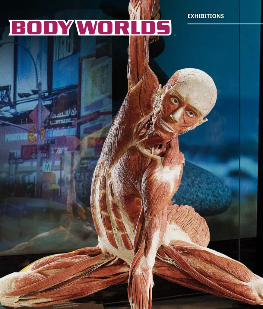
FR:EIA — Fascia Revealed: Educating Interconnected Anatomy — is the first attempt of its kind in the world.
This article sets out what project FR:EIA came from, what it makes visible, and also what it does not make visible, from the perspective of the body’s [science]. Avoiding excessive mythologizing, I want to lay out both the significance and the limits of this specimen.
The Technique of Plastination
This is a method developed by the anatomist and physician Gunther von Hagens, in which the water and fat contained in living tissue are replaced with polymer, producing a specimen that neither decays nor dries out and can be preserved semi-permanently. The BODY WORLDS exhibitions of human specimens that von Hagens has staged around the world rest on this technique.
The advantage of plastination is that it fixes the three-dimensional structure of tissue as it is. That property made possible something conventional dissection could not do: showing the network of fascia running continuously through the whole body, in three dimensions, without breaking it apart.
The Fascial Net Plastination Project (FNPP)
FR:EIA came out of the Fascial Net Plastination Project (FNPP).
The project was launched in 2018 by Robert Schleip, a Rolfer and a leading figure in fascia research. It proceeded as an interdisciplinary collaboration between the technical team at BODY WORLDS and the Fascia Research Society. Specialists in anatomy, dissection technique, and plastination gathered internationally and spent roughly three years on an ambitious aim: to show the fascial network of the whole body, three-dimensionally, as a single specimen.
What that work produced was the world’s first whole-body 3D fascia plastinate, FR:EIA. The name comes from the initials of Fascia Revealed: Educating Interconnected Anatomy.
“Removing Fascia in Order to Show Fascia”
The most interesting thing about how FR:EIA was made is the paradox at the root of it.
Gary Carter, the anatomist who served as design and co-dissection lead on the team, heads off an important misunderstanding about the specimen. Hearing “the fascia of the whole body,” most people picture a body covered entirely in a honeycomb mesh. FR:EIA is not that, he says.
If all the fascia of the body were left in place, it would simply be a mass of white membrane, and nothing would be visible at all. So the team decided, one by one, which of the fascia normally removed in dissection to keep and which to take away. Removing fascia in order to show fascia — this succession of choices is what gave the specimen its fascial focus.
Many specialists were involved in the making. The project proceeded under the direction of Robert Schleip and Carla Stecco, with Vladimir Chereminskiy as chief anatomist on the Body Worlds side and Rachelle Clauson, FNPP’s Director of Creative and Administrative Affairs, overseeing creative and administrative matters. Around them, a volunteer dissection team of some forty people took part.
Unveiling — From an Online Premiere to Montreal, and Then Berlin
FR:EIA was first shown to the world on November 24, 2021, in an online premiere watched by more than a thousand participants. The following year, in September 2022, the specimen itself was presented in person for the first time at the 6th International Fascia Research Congress in Montreal, Canada. A specimen in which connective tissue can be seen three-dimensionally, as it is, was received by physicians, therapists, and fascia researchers as a research object without parallel.
For Rolfing — Structural Integration — this unveiling carried a further meaning. It is fair to say that the view Ida Rolf had been setting out since the 1950s, with fascia at its center, that the whole body is connected, was given visual support. Schleip has pointed out that working with fascia can have effects not only locally but throughout the body, because fascia is interrelated and connects everything, and has said that FR:EIA gave this idea a renewed contemporary significance.
The work was carried out at von Hagens’s facility, the Plastinarium, in Guben, Germany, and the completed specimen is now on permanent display at the BODY WORLDS museum in Berlin. In response to growing public interest in fascia research, the museum has established a dedicated area for fascia plastinates, with FR:EIA at its center.
The European Rolfing Association (ERA) has entered into a cooperative relationship with the BODY WORLDS museum in Berlin in order to convey the significance of this fascia research more widely.
Why Three Years? — What Makes This Attempt Interesting
To keep FR:EIA from ending as a curiosity, there is a question worth pressing on here. Why did an international team spend three years making it? And what was the value in that?
For Some Five Hundred Years, Anatomy Threw Fascia Away
As noted at the outset, fascia was long discarded in the dissection room. But that was not mere carelessness. Since Vesalius in the Renaissance, modern anatomy developed with “cleaning the specimen” as its craft — in order to observe the form of muscles and organs accurately, removing the membranes covering them was, if anything, the correct procedure. Modern anatomy, in other words, had built a blind spot into its own method: not seeing fascia.
What makes FR:EIA interesting is not that it is a display object. It is that it puts a question to a way of seeing the body that has held for some five hundred years. A tissue that had been cleaned off and thrown away was placed, for the first time, at the center of a specimen. This is a story about a technique and, at the same time, about a shift in how the human body is regarded.
What Cannot Be Seen Cannot Be Studied
The claim that fascia matters had been made empirically by many clinicians, Ida Rolf among them. But that claim had a decisive weakness: there was no means of showing the whole of it.
It is like speaking about a continent for which no map exists. However much one says “it is there” and “it is important,” without an object anyone can examine, the discussion floats free. Much of those three years went into this single point: making the invisible visible.
Why It Is So Difficult
Preserving the fascia of a whole body as a single specimen is technically very hard.
One problem is fat. Fat cannot be preserved and must be dissolved out, but the superficial layers of fascia hold fat within their honeycomb structure like honey. Draw out the honey, and the comb collapses. Keeping that delicate structure intact, still full, was itself a difficult problem.
Beyond that, the dissection and fixation had to preserve the continuity of the whole body. There is no reassembling afterward. On a single donated body, it is a succession of irreversible judgments — once cut, never restored. Those three years accumulated under the tension of a single attempt.
What to Keep and What to Remove — The Judgments Are the Content
The choice touched on earlier, removing fascia in order to show fascia, is in fact where the most essential difficulty lies, and at the same time it is the content of this specimen.
What to keep, what to take away. Through countless such judgments, the makers decided, one at a time, what matters about fascia. FR:EIA is therefore not a neutral photograph of reality as it is. It embodies an interpretation and an argument, as an object. The gaze of its makers is inscribed in it. That is why it is not merely a preserved thing but an intellectual work.
The Value — Turning a Metaphor into an Object
So what did those three years produce?
Before FR:EIA, the phrase “everything is connected through fascia” was a clinical intuition, and at times sounded like a slogan. After FR:EIA, it became an object in front of you, one you can walk all the way around. A claim that had lived in the territory of belief and metaphor was moved into a shared object anyone can examine.
This is not the discovery of a new fact. But in the same sense that Vesalius did not discover a new organ and instead changed how the body was seen, FR:EIA also changed a way of seeing. It gave orthopedics, physiotherapy, osteopathy, and bodywork — fields that had each spoken about fascia separately — a single reference point they can all point to. It pushed fascia from background and packing material to the leading role. Schleip’s description of FR:EIA as a historical extension of conventional anatomy presumably reflects the size of that shift.
The value of those three years does not lie in any dramatic proof of effect. What had not been visible became visible, and with it the ground of the conversation about fascia changed. That shift is the most interesting thing about the attempt.
What the Curator Saw — Angelina Whalley on Making FR:EIA
On how FR:EIA was made, there is a valuable primary source: an interview with Angelina Whalley, physician and curator at Body Worlds, published in Structure, Function, Integration, the journal of the Dr. Ida Rolf Institute (July 2022). She is an anatomist who has worked alongside von Hagens and has conceived and designed the Body Worlds exhibitions, work that has reached more than 52 million people.
What strikes first is Whalley’s own frank admission. Like the anatomists of the past two centuries, the Body Worlds team had also been cutting fascia away and discarding it as dissection waste. That stance changed when Schleip, the fascia researcher, brought the project to them. The theme this article has been describing — fascia as the discarded tissue — is confirmed by the makers themselves.
Whalley conveys the difficulty of the work through a single comparison. Think of peanut brittle: the nuts are the bones and muscles, and the caramel that binds them is the fascia. If you want to show the caramel, the only way to see inside is to break away what surrounds it. Here again the paradox surfaces: breaking in order to show.
The scale was not ordinary either. The superficial layers of fascia, rich in fat, were particularly difficult to handle; dissection by the Body Worlds team alone took 800 hours, and volunteers from the Fascia Research Society spent a further 3,000. Three years was the accumulation of that vast amount of handwork.
The final posture has a story as well. The original plan had the right arm raised, holding a heart aloft. But it also looked somewhat as though the heart were being thrown away, and in a specimen meant to focus on fascia, the heart would have taken the leading role. What was chosen in the end was a dancing posture with the arms lowered — a pose suited to a fascia specimen, reflecting extension, suppleness, and movement.
Of particular interest is the experience built into the Berlin exhibit.
Visitors first bend forward. Next, they work the fascia on the soles of both feet with a foam roller. Then they bend forward again. Many are surprised to find their hands reaching further than before — though all they touched was the soles of the feet. It is a device that lets the continuity of fascia through the whole body be felt in the body rather than understood in the head. The hall also holds a tensegrity model, showing that bones are not stacked in compression against one another but float within lines of fascial tension.
Whalley describes the significance of the exhibit this way: musculoskeletal pain does not necessarily come from the joints; it can come from fascia. That knowledge is what changes how a person meets and uses their body. Not only specialists but many people, including those who come to fascia through yoga, stand looking at this specimen.
What This Method Reveals — Three Things Fascia Shows Us
FR:EIA is not a device that produces dramatic new discoveries. It is more accurate to see it as giving an unprecedented view of structures already known anatomically. But that new view does contain information hard to capture by other methods. What fixing the fascia of a whole body in three dimensions reveals is mainly the following three things.
1. The Body Is Connected Without Interruption
In conventional dissection, fascia is removed in order to make muscles and organs easier to see. At that moment, the connection fascia held is lost. Imaging such as ultrasound and MRI, on the other hand, can show living tissue but only a given cross-section or region; it cannot capture the continuity of the whole body in a single image.
What FR:EIA showed fills the space between them. From head to toes, fascia is preserved three-dimensionally as one network continuing without interruption. That fascia runs connected through the body is something many practitioners have described empirically; FR:EIA is what made it visible as a single specimen.
2. Fascia Becomes a Path for Transmitting Force
Being connected means that force applied in one place can travel to a place far from it. Recent research has shown that force is transmitted between muscles by way of fascia — myofascial force transmission. The work of Peter Huijing, who is also an academic advisor to the ERA, on force transmission in skeletal muscle is one representative line of this. Even in whole-body movement such as walking, the handing along of tension through fascia is thought to support the movement.
FR:EIA makes visible, as continuous tissue, the paths along which force can travel. Why tension in the calf cannot be unrelated to the state of the back or the neck — the specimen shows the anatomical ground for that.
3. But Structure Alone Is Not Enough — the Properties of Living Fascia
Here a distinction should be made honestly. What FR:EIA shows is a fixed, motionless, post-mortem structure. Yet much of what makes fascia an interesting tissue for bodywork lies in properties that appear only in a living body. Those have been revealed not by FR:EIA but by other bodies of research.
Schleip and colleagues, for instance, have pointed out that fascia may be one of the body’s richest sensory organs, containing an enormous number of nerve endings. The hypothesis has also been put forward that fascia contains myofibroblasts and may contract slowly, like smooth muscle. Fascia is further thought to be closely involved with the autonomic nervous system.
The whole picture, in other words, comes into view only when the map of structure that FR:EIA shows is laid over the research dealing with the function of living fascia. To think about what may happen when hands-on work engages the body, neither structure alone nor function alone is enough. Both are needed.
The Relationship to Rolfing and Bodywork
Ida Rolf’s Starting Point
Ida Rolf, the originator of Rolfing, began from the idea that the body is organized by fascia and that its structure can be reorganized within gravity. Working carefully with fascia that has hardened and shortened, and bringing the balance of the whole body into order in its relationship with gravity — this is the notion at the root of Rolfing. Rolf is said to have stated repeatedly, in substance, that everything is connected through fascia.
FR:EIA gives that starting point a visible ground. A body appearing not as an assemblage of separate parts but as one continuous network of membrane resonates directly with the view of the body Rolfing has taken as its premise.
Anatomy Trains — a Map Born out of Rolfing
The practitioner who organized this view of the body into a more systematic map is Tom Myers, with Anatomy Trains. Myers studied Structural Integration under Ida Rolf, and developed the concept while teaching fascial anatomy at the Rolf Institute in the 1990s. The starting point was an attempt to draw Rolf’s claim that everything is connected through fascia as specific linkages.
Anatomy Trains sets out several myofascial meridians running through the body. Taking the Superficial Back Line, which runs vertically up the back of the body, as a single line, for instance, brings into view a whole picture of posture and movement that looking at the hamstrings alone cannot give. This line running from Rolf to Myers tells a good deal about how Rolfing and bodywork have understood connection through the whole body.
Why Rolfing Works with the Whole Body, in a Sequence
If fascia is continuous, then a restriction in one place produces a pull in a place far from it. So Rolfing does not knead only the painful spot; it works with the whole body in sequence through a series of ten sessions. The structure of the Ten Series, moving in stages from superficial fascia toward the deeper layers, corresponds to the fact that fascia is layered.
The ground FR:EIA made visible — fascia as continuous and layered — helps in understanding why Rolfing proceeds in the shape it does.
What Can Be Said, and What Cannot — an Honest Boundary
Having drawn the connection between fascia and Rolfing this far, a boundary should be drawn clearly so as not to overreach.
First, FR:EIA was made from a single donated body and is one specimen; it does not represent a statistical population. And what it shows is fixed structure — not the glide or plasticity of living tissue, still less proof of whether fascia changes through hands-on work, or of clinical effect. Visualization is visualization, not proof of effect.
Second, the continuity of fascia — the myofascial meridians of Anatomy Trains — stands at a stage where some has been supported by dissection research and some remains unconfirmed.
In the verification work of Jan Wilke and colleagues, while some lines such as the Superficial Back Line were supported, there are also parts, such as the Superficial Front Line, where anatomical linkage has not been confirmed. Myers himself presents Anatomy Trains not as established fact but as one map that may be useful. FR:EIA makes continuity visible, but it does not verify every proposed line.
What FR:EIA and fascia research do show, then, is the soundness of a premise: that fascia is an independent, continuous, sensation-rich tissue worth taking seriously. It shows that what Rolfing works with is a real and definite tissue. It does not, on the other hand, prove that Rolfing works. Verification of effect is a separate research question, one taken up by Katja Bartsch, Robert Schleip, and others.
Once the Technique Is Established, Where Does This Go?
As we have seen, the greatest difficulty with FR:EIA lay in the technique itself: preserving fascia alone without its collapsing. Similar attempts had in fact been made before, but the fat could not be removed successfully and each was shelved. FNPP began with small partial specimens and confirmed, one at a time, how plastination behaves when fascia alone is isolated, precisely in order to establish this technique.
Once that technique is established and reproducible, fascia research may widen in several directions.
1. From One to Many — the Perspective of Comparison
FR:EIA at present is, so to speak, a single sample (n = 1). But if the technique stabilizes, several bodies can be made into specimens by the same method. Differences in age, sex, and exercise history; bodies carrying chronic pain or postural bias, and bodies not carrying them — the fascial structures of these could be set side by side and compared. The question could advance from the general “fascia is connected” to the specific: in what kind of body, and where, does it differ, and how?
2. Becoming a Common Language for Education
This movement has already begun. The collection of partial specimens produced by FNPP was placed on long-term loan to the University of Padua in Italy in 2023, and is used in the anatomical education of medical students. Padua is the university where Carla Stecco, a leading figure in fascial anatomy and an anatomical advisor on FR:EIA, is based.
Once, seeing fascia meant attending one of a limited number of dissection courses. A specimen that does not break down, does not smell, and lasts semi-permanently opens that experience to educational settings worldwide. Fascia can become a common language, something everyone can learn while pointing at the same object, rather than tacit knowledge held by specialists alone.
3. Connecting with Images of the Living Body
A plastinate does not move, but in exchange it holds a three-dimensional “correct answer” of the anatomy. This makes it a valuable reference point for in vivo imaging such as ultrasound and MRI. What structures an indistinct image taken in a living body is actually showing can be read with greater precision by comparison against a fixed specimen. This is one point at which research such as Katja Bartsch’s assessment of fascial mobility by ultrasound connects.
Structure (the plastinate), living structure (imaging), and function (research on mechanics and the nervous system) — only when these three mesh does the whole picture of fascia as a tissue come closer. Establishing the technique is the work of making the first of those legs more solid.
Limits That Do Not Change
However refined the technique becomes, the fact noted earlier — that a plastinate is a fixed, post-mortem structure — does not change. It does not become an all-purpose method; it remains one perspective, meaningful only in combination with research on living bodies.
Conclusion
FR:EIA is neither dramatic proof of effect nor all-purpose evidence. What one specimen shows is a stance — looking at the tissue called fascia head-on — and a definite ground: that it is a real continuum. A tissue long cleaned off and discarded was placed, for the first time, at the center of a whole-body specimen. That shift is itself emblematic of how the field of fascia research has developed.
Fascia, what Rolfing works with, is now displayed in a museum as an independent object of research and education. Granting that the premise is sound, what actually happens in your own body is something to be confirmed not by a judgment in the head but by the body itself. This field, in a breadth that a single word like “pseudoscience” cannot capture, is still moving forward.
Reference Links
- BODY WORLDS: FR:EIA
- Whalley A, Hack LA. Fascia Insights: Curating FR:EIA, the First Full-Body Fascia-Oriented Plastination Exhibit.Structure, Function, Integration: The Journal of the Dr. Ida Rolf Institute. 2022;50(2):4–9.
- Fascial Net Plastination Project(Wikipedia)
- Fascial Net Plastination Project official YouTube channel
- Fascia Research Society
- Tom Myers, Anatomy Trains: Myofascial Meridians(4th ed., Elsevier)
- European Rolfing Association: Scientific Research
- Ida P. Rolf Research Foundation
Related Articles
- Is Rolfing Pseudoscience? — Understanding Rolfing Through the Science of the Body
- Twenty-Three Years of Records, Brought to Light by a Leading Fascia Researcher — From a New Study of the Rolfing Ten-Series
- Rolfing Is Studied in University Laboratories — Doctoral Research on Fascia at the Technical University of Munich
Hidefumi Otsuka, Ph.D. | Certified Advanced Rolfer™ / Rolf Movement Practitioner
Completed his doctorate at the University of Tokyo Graduate School of Medicine. After eleven years in medical marketing at a foreign pharmaceutical company, he has offered Rolfing® sessions in Shibuya since 2015. His work centers on the integration of science and practice.